"Discuss the importance of the Frank-Starling mechanism and discuss the balancing between the cardiac and vascular function curves."
Frank-Starling mechanism
Starling's law of the heart is the culmination of the work of two gentlemen. Starling’s law states that "energy of contraction is proportional to the initial length of the cardiac muscle fibre". This conclusion was reached from work done by Frank in 1895 and from work done by Starling himself in 1914.
Frank had performed experiments on isolated frog hearts where he had found that increasing the force on the myocardial fibres (the preload) increased the force of contraction.
Starling performed a more complex experiment whereby he was able to adjust the venous pressure (the pre-load), and the peripheral resistance. Rubber tubing was used to transfer the blood from the aorta into a venous reservoir. From the reservoir, blood was returned to the heart through a clamped piece of tubing. This allowed the venous return pressure to be changed by either raising or lowering the reservoir or opening the clamp. The peripheral resistance could also be varied through a pressure limiting system installed in the rubber tubing known as a Starling resistance. The cardiac output was measured by measuring the outflow of blood into the venous reservoir. Other measurements were taken of arterial pressure, venous pressure at the right atrium, pressure at the left atrium and the volume of the ventricle. The lungs were left attached to maintain the pulmonary blood flow system.
With this arrangement and the denervated heart beating steadily Starling carried out his experiment.
It was found that increasing the right atrial pressure caused, for a few beats, a progressive increase in the ventricular volume until a new steady state was reached. A similar pattern was found on increasing the peripheral resistance.
The indication from this was that in the first instance the increased venous pressure caused an increased flow of blood into the ventricles. This led to a disparity between the cardiac output and the influx. As the ventricles expanded to accommodate the extra volume of blood the myocardial fibres became stretched. As the fibres stretched, they produced more force and a larger volume of blood was expelled from the heart in systole.
In the latter case an increase in peripheral resistance reduced the volume of blood that was able to leave the heart. The ventricles again, became distended as they accumulated the blood that they were unable to pump away. After several beats and an increase in ventricular volume the cardiac output returned to normal.
The increased length of the myocardial fibres is thought to provide additional contractile force through an increased availability of myofilament crossbridges. There may also be an effect in altering the sensitivity to calcium.
Physiologically the Frank-Starling mechanism is important in balancing the output of the two ventricles of the heart. Any increase in the output of the right ventricle will lead to an increased inflow to the left ventricle. Were this not able to react by increasing its output there would be a steady increase in its volume until something catastrophic occurs. It is to be noted that the Frank-Starling curve has a "descending limb" at high filling volumes. In this region further stretching of the cardiac fibres leads to a break down in their structural integrity and subsequent drop in cardiac output. A catastrophe would occur were this state ever to occur in the human body. The diminished cardiac output would be unable to pump away all the blood that fills the ventricle during diastole. As the blood accumulates, the cardiac output falls still further until cardiac failure occurred.
The changes in force of contraction can also help to regulate cardiac output in alteration of heart rate. If the heart were to slow then there would the increased time for diastole leads to increased ventricular filling. The increased volume of the ventricle would d increase its’ output. Thus the cardiac output can be maintained in a fairly constant state.
In exercise the cardiac output needs to be increased dramatically and there is a definite correlation between increased heart rate and increased cardiac output. It is now thought that the increased heart rate is not the primary cause of increased cardiac output but rather a factor that permits it. The lowering of the resistance of the vasculature is thought to play the major role.
Vascular function curve
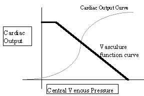
Often mathematical functions can be use to describe system characteristics. The cardiovascular system is no exception. There are a multitude of functions to describe virtually all behaviours of the system. Two of concern here are known as the cardiac function curve and the vascular function curve. They are both graphs representing cardiac output plotted against central venous pressure.
The cardiac function curve is essentially a restatement of the Frank Starling law. It is plotted as the cardiac output dependent on the venous pressure and is a function solely of the heart. The vascular function curve is the reverse of this in that it sows the venous pressure dependent on the cardiac output and is a function of the vasculature.
The vascular function curve shows an inverse relationship between central venous pressure and cardiac output. This is explained in the following manner: should there be a sudden increase in cardiac output the arterial pressure will rise as the flow through the peripheral resistance can only increase once this has happened. As the heart is pumping more blood out of the venous system, the pressure here will be lowered.
The pressure for which cardiac output is zero (fro example, in cardiac arrest) is termed the mean circulatory pressure. Whilst the vascular system remains closed and in a passive state, this will remain constant. It will be noted that as the cardiac output increases, the central venous pressure does not simply pass through the origin and become negative. At a point equal to a cardiac output of about 7 L min-1, the venous pressure suddenly drops away and no further increase in cardiac output is possible. At this point the veins have dropped below ambient pressure. The flexible wall of the veins collapses in and prevents flow of blood into the heart.
Balancing of cardiac and vascular curves
Because of the cardiovascular system is essentially tightly closed, a change in one variable will have to affect the other, which will then affect the first, etc. Any feedback system like that can operate as a negative or positive feedback loop. It can clearly be seen that if positive feedback occurred then a state of cardiac failure would exist; either through too great a depression of the cardiac output or from an overloading of the heart and subsequent rupture. Thus, the two function curves act on each other to drive the system to an equilibrium value.
A representation of the two curves. By convention the vascular function curve is normally drawn with cardiac output as the abscissa. They have been swapped here to allow its’ overlay on the cardiac output curve.
A suddenly increased cardiac output would involve a transient decrease in the volume of blood in the central venous system. The blood flowing into the veins through the peripheral resistance would be less than that being pumped away by the heart. This causes a lowering of central venous pressure. The lower venous pressure will reduce the amount of blood flowing into the ventricles in diastole. By Starlings Law, this will reduce the output of the heart. Hence the reduction in venous pressure will be reduced, thus the increases in cardiac output will be reduced, etc. etc..
Clearly there can be no big difference in the long term between the two functions of venous return and cardiac output. A heart cannot pump more blood out of the venous system than there is delivered to it. The venous return pressure and the cardiac must balance themselves against each other to maintain the viability of the system.
The body as a dynamic system
For the body to be properly supplied by blood the cardiovascular system needs to adjust to its changing needs. The cardiac and vascular function curves enable us to understand some of the responses of the system to change. Three things that are often changed are peripheral resistance, myocardial contractility and blood volume.
To consider the effects of such changes it is helpful to be aware of the changes brought about on the curves themselves. The cardiovascular system as stated earlier will always tend to drive itself towards the cross over of the two functions.
Peripheral resistance is a variable that changes the vascular function curve and the cardiac function curve. As peripheral resistance increases, the cardiac function curve moves downward. At any given venous pressure the heart is able to pump less blood against the increased pressure. The vascular curve on the other hand undergoes a rotation. The mean circulatory pressure will be unaffected by the change as at zero cardiac output the pressure is reliant solely on the contractility of the blood vessels and the blood volume in them. However, an increase in peripheral resistance will increase the arterial pressure needed to be overcome to produce any given cardiac output. As a result the effects of its change are not particularly easy to predict. Prediction relies on establishing the relative magnitude of the shifts of both curves. If the vascular curve shifts more than the cardiac then the equilibrium point will fall down and to the left. Both venous pressure and cardiac output will fall. Were the cardiac function curve to shift more than the vascular then the equilibrium point would shift down and to the right of its initial position. That would mean that cardiac output would drop but venous pressure would actually rise slightly.
Myocardial contractility is another factor that often changes in the body. For example, stimulation by the sympathetic nervous system can cause an increase in ventricular contractility. Increased contractility leads to a raising of the cardiac function curve (a given venous pressure produces a greater cardiac output). On initial stimulation by the sympathetic nerves, the increased contractility produces an abruptly increased cardiac output. Venous pressure would then gradually fall due to the increased transfer of blood from the venous to the arterial side of the heart. The fall of venous pressure reduces the cardiac level and so on until a new equilibrium is reached. This equilibrium is at a higher cardiac output and lower venous pressure than without sympathetic stimulation.
The third major factor and one that is often clinically relevant is the reaction of the cardiovascular system to changes in blood volume (as in haemorrhage or blood transfusion). Because the circulation is effectively a closed system any change in blood volume is going to affect the venous pressure. Thus will not affect the cardiac function curve but will mean that the vascular function curve is shifted up. (A given venous pressure produces a higher cardiac output.) Thus the equilibrium point is shifted upwards on the cardiac function curve. Both cardiac output and central venous pressure will be increased.
(c)1998 Nick Manville